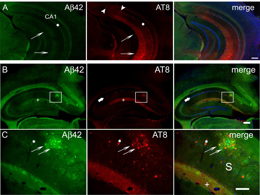
Figure 2. Aβ42 accumulation and tau hyperphosphorylation co-occur in the SLM of the CA1 region of hippocampus in 3xTg-AD mouse brain.
(A) Early Aβ42 accumulation and tau hyperphosphorylation in 5-month-old 3xTg-AD mouse brain prior to plaques and tangles. In the CA1 region of the hippocampus, Aβ42 is prominent in the pyramidal cell layer (asterisk) and in the SLM layer (thin arrows). AT8 antibody labeling of hyperphosphorylated tau is most pronounced in the SLM layer (thin arrows). A few pyramidal neurons with AT8 labeling (arrowheads) are evident in the subiculum at this age. (B) Section through the rostral hippocampus from a 13-month-old 3xTg-AD mouse labeled with Aβ42 and AT8 antibodies by immunofluorescence. Nuclei were labeled with Hoechst dye 33342 to distinguish the subiculum from the hippocampal CA1 region, where the pyramidal cell nuclei are clearly lined up. Amyloid plaques were apparent within the subiculum of the hippocampus (boxed area), but were also evident in the SLM of CA1. Both Aβ42 peptides and hyperphosphorylated tau were markedly increased in the SLM (plus signs) of CA1. Hyperphosphorylated tau (AT8) also labeled a few cell bodies within CA1 (thick arrows). (C) Higher magnification image of the subiculum from the squared area in (B) with prominent plaque pathology. Aβ42 and hyperphosphorylated tau co-localized in several pyramidal cell bodies (asterisks). Hyperphosphorylated tau and Aβ42 peptide co-localization was prominent in dystrophic neurites around a large amyloid plaque (thin arrows). Punctate co-localization of Aβ42 and hyperphosphorylated tau was also evident in the SLM (plus signs). Abbreviation: S, subiculum, SLM, stratum lacunosum-moleculare. Scale bars: 250 µm (A, B); 100 µm (C).

