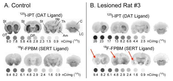
FIGURE 5.
[125I]IPT and [18F]FPBM binding in the striatum of the three Parkinson’s model rats were examined through in vitro autoradiography. (A) In the control rat, the dopamine transporter ligand, [125I]IPT, strongly binds in both the left and right striatum, as expected. [18F]FPBM also appears to have equal amounts of binding in the left and right striatum. Binding of [18F]FPBM appears less intense than [125I]IPT due to the natural lower level of serotonergic innervation compared to dopaminergic innervation in the striatum. (B) [125I]IPT binding in the left striatum is clearly reduced in rat #3. Rat #3 also had visible differences in [18F]FPBM binding between the left (red arrows) and right striatum. Rat #1 and #2 also showed reduced [125I]IPT binding in the left striatum but did not show any differences with [18F]FPBM (not shown). 125I standards were exposed to the film along with the brain sections. DAT: dopamine transporter, SERT: serotonin transporter, St: striatum, OT: olfactory tubercle, GP: globus pallidus, Hp: hippocampus, Th: thalamus, Am: amygdala, Cb: cerebellum, LC: locus coeruleus (Please note: 1nCi = 0.037 Bq).

