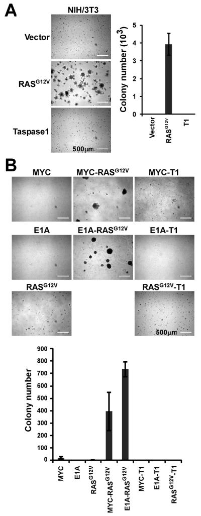
Figure 2.
Taspase1 is not a Classical Oncogene. A, NIH/3T3 cells were transduced with the indicated genes and plated on soft agar. 5×10⁴ cells were plated in each 6 cm dish. Positive clones (>200 μm) were scored 10 days after the initial plating. Data presented are mean ± SD of duplicates of two independent experiments. B, Wild-type primary MEFs were transduced with the indicated pairs of genes and plated on soft agar. 5×10⁴ cells were plated in each 6 cm dish. Positive clones (>200 μm) were scored 2-3 weeks after the initial plating. Data presented are mean ± SD of triplicates of two independent experiments. Inserts are higher-magnification images. Scale bar equals 500 μm. T1 denotes Taspase1.

