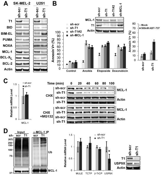
Figure 5.
Taspase1 Deficiency Leads to Decreased MCL-1 Protein and Increased Cancer Cell Death upon Treatment with Chemotherapeutic Agents and ABT-737. A, Cellular extracts of SK-MEL-2 or U251 cells with control- or Taspase1-knockdown were subjected to Western blot analyses using the indicated antibodies. B, U251 cells with the indicated knockdown were treated with various apoptotic stimuli, stained with annexin-V, and analyzed by FACS. For anoikis assays, cells were plated in PolyHEMA coated plates for 3 days. For chemotherapeutic agent-induced cell death, cells were treated with 100 μg/mL of etoposide or 10 μM of doxorubicin for 30 hours. Data presented are mean ± SD of duplicates of two independent experiments. U251 cells with indicated knockdown were mock (DMSO) or ABT-737 treated for 24 hours, stained with annexin-V, and analyzed by FACS. Data presented are mean ± SD of triplicates of two independent experiments. C, The transcript levels of MCL-1 in control- or Taspase1-knockdown U251 cells were determined by quantitative RT-PCR analysis (left). U251 cells with indicated knockdown were subjected to cyclohexamide or cyclohexamide plus MG132 treatment for the indicated periods of time, and the protein levels of MCL-1 and β-actin were determined by immunoblot (right). D, The ubiquitination status of MCL-1 in U251 cells with control- or Taspase1-knockdown were treated with MG132, and subjected to anti-MCL-1 immunoprecipitation followed by anti-ubiquitin immunoblots. Transcript levels of known MCL-1 stability regulators were analyzed and the protein expression of USP9X was determined. Asterisk indicates P < 0.01. Data presented in C and D are mean ± SD of duplicates of three independent experiments and the transcript level in control-knockdown cells was assigned as 1.

