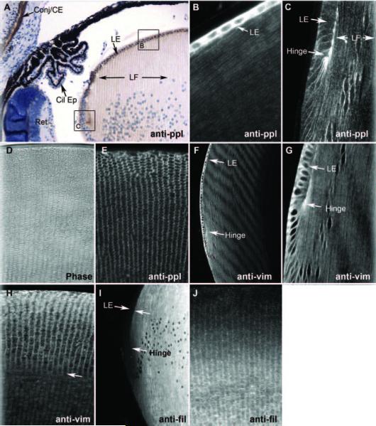
Figure 4.
Immunocytochemistry. Six-micron-thick paraffin sections probed with antibodies to (A) periplakin, then localized with HRP-goat anti-rabbit antibodies. Brown: reaction product can be seen in the corneal epithelium and conjunctiva (conj/CE) but not in ciliary epithelium (Cil Ep) or retina (Ret). The lens epithelium (LE) is very heavily reacted, as is the “hinge” region. Labeling of fiber cells (LF) is principally at the membrane. Areas comparable to boxed areas “B” and “C” are shown in (B) and (C). (B) Periplakin immunofluorescence labeling of more anterior epithelium and underlying fiber cells. Epithelial reactivity is very high, but fiber cell membrane labeling is also evident. (C) Periplakin labeling of lens “bow” or equatorial region, where fiber cell elongation begins. Epithelial cytoplasmic labeling is sharply reduced, and most labeling is at the plasma membrane. (D) Phase-contrast image to demonstrate histology of a coronal section of mid-lens. (E) Coronal section. Periplakin immunofluorescence showing cross-sectioned fiber cells. (F-H) Vimentin immunofluorescence of sagittal (F, G) and coronal (G) sections. (I, J) Filensin immunofluorescence in sagittal (I) and coronal (J) sections. (I) Note the complete absence of the labeling of lens epithelium.

