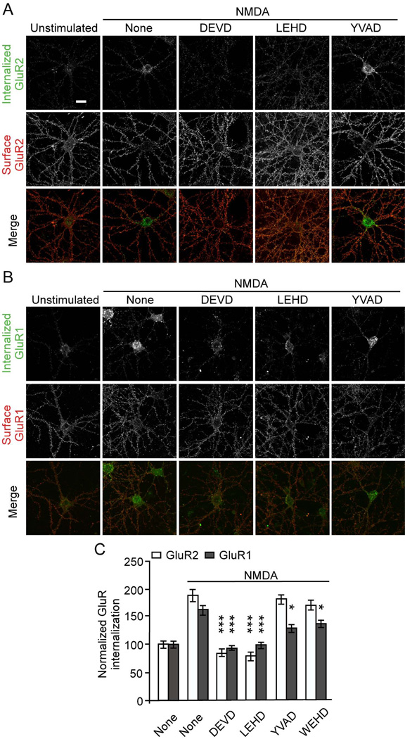
Figure 4.
Caspase inhibitors DEVD- and LEHD-FMK block NMDA-induced AMPA receptor internalization. Antibody-feeding internalization assay for endogenous GluR2 (A) and GluR1 (B) in hippocampal neurons stimulated with NMDA (50 µM for 10 min). Neurons were pretreated with various caspase inhibitors (5 µM) as indicated. A and B show double-label immunostaining for internalized GluR (green, first row) and surface-remaining GluR (red, second row), and merge (in color, third row). Individual channels are shown in grayscale. Quantitation in C shows internalization index (integrated fluorescence intensity of internalized GluR / integrated fluorescence intensity of internalized GluR plus surface GluR) normalized to unstimulated untreated cells (Lee et al., 2002). For all quantitations, n=15 neurons for each group. *p<0.05, ***p<0.001, compared to NMDA treated cells transfected with µ-gal. The graph shows mean ± SEM. Scale bar, 20 µm. See also Figure S3.

