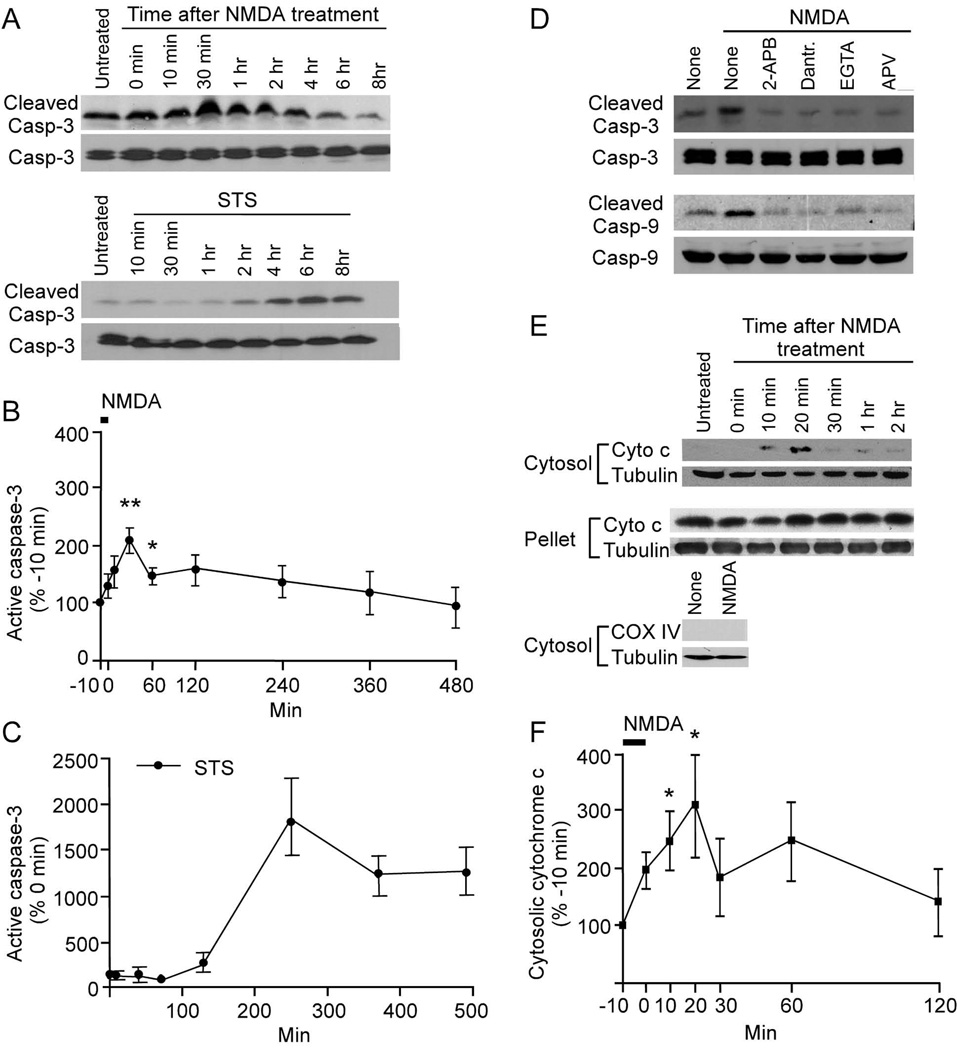
Figure 6.
Transient activation of caspase-3 in neurons by NMDA. (A, D, E) Immunoblots showing cleaved (active) caspase-3 and caspase-9, and cytosolic cytochrome c in cortical neuronal cultures (DIV18) at various times after treatment with NMDA (50 µM for 10 min) or staurosporine (STS, 1 µM throughout the experimental period). In (D), neurons were preincubated as indicated with 2-APB (30 µM), dantrolene (100 µM), DL-APV (100 µM) or EGTA (5 mM) for 15 min before NMDA treatment, and were immunoblotted 30 min after NMDA stimulation for active (cleaved) and total caspase-3,-9; and cytochrome c in the cytosolic and pellet fractions. (B, C, F) Quantitation (mean ± SEM) of immunoblot band intensities normalized to values before treatment (NMDA was applied from −10 to 0 min; STS treatment began at 0 min); n=3 for each time point. See also Figure S5.

