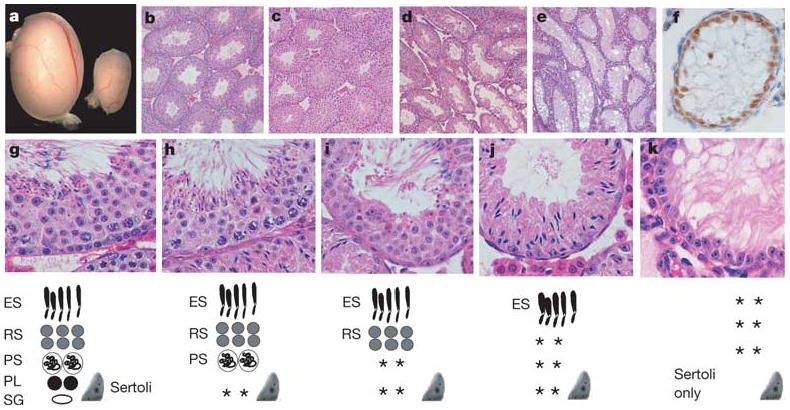
Figure 1. Spermatogonial depletion and Sertoli-cell-only syndrome in ERM−/− mice.

a, Ten-week wild-type (left) and ERM−/− (right) testes. b, Wild-type seminiferous tubules. c–e, Progressive germ-cell loss in ERM−/− testis at 4 weeks (c), 6 weeks (d) and 10 weeks (e). f, Persistent Sertoli cells in ERM−/− tubules indicated by GATA-1 expression at 10 weeks. g–k, Normal spermatogenesis with spermatogonial depletion. Histology of wild-type (g) or ERM−/− (h–k) testes at 6 weeks of age. Diagrams of seminiferous epithelium are shown at the bottom: ES, elongated spermatid; RS, round spermatid; PS, pachytene spermatocyte; PL, preleptotene spermatocyte; Sg, spermatogonium; Sertoli, Sertoli cell; asterisks, missing cell populations.
