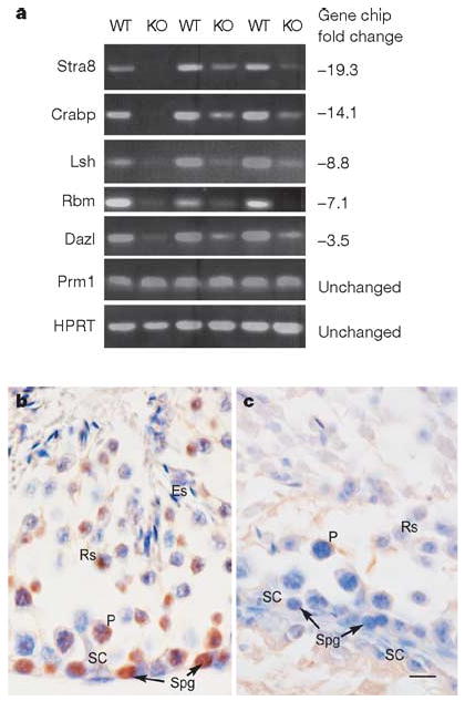
Figure 2. Selective reduction of spermatogonia-specific genes in ERM−/− testes.

a, RT–PCR analyses of three independent pairs of wild-type (WT) and ERM−/− (KO) male littermates are shown for the indicated genes. Numbers at the right represent the relative fold reduction measured by microarray analysis (Supplementary Table 1). HPRT, hypoxanthine–guanine phosphoribosyltransferase. b, c, Expression of Plzf in 6-week-old wild-type (b) and ERM−/− (c) testes by immunohistochemistry. Cells indicated are as follows: ES, elongated spermatid; RS, round spermatid; PS, pachytene spermatocyte; Spg, spermatogonium; Sc, Sertoli cell. Scale bar, 10 μm.
