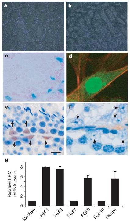
Figure 3. ERM expression in testis is restricted to Sertoli cells.

a, b, In situ hybridization for ERM mRNA in 10-week wild-type (a) and ERM−/− (b) testes. c, Testes from 6-week ERMIRES-LacZ heterozygous males were stained for LacZ expression. d, TM4 Sertoli cells were infected with ERM-GFP-RV retrovirus and analysed by confocal microscopy. e, f, Immunolocalization of ERM protein in adult wild-type (e) and ERM−/− (f) testis with anti-ERM monoclonal antibody. Arrows indicate nuclei of Sertoli cells in wild-type (e) and ERM−/− (f) testes. g, ERM mRNA expression in TM4 cells after treatment with various FGFs. Data are fold induction (means + s.d. for two independent measurements) compared with ERM expression in medium.
