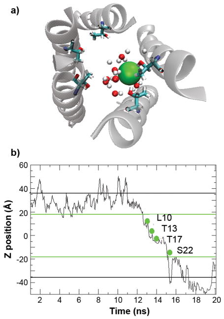
Figure 9.
a) A snapshot that shows the interactions between Cl− (shown as the green sphere) and water molecules and Thr17 hydroxyl side chains inside the channel. Only those water molecules located within 5 Å of chloride are shown. b) Position of a Cl−along the membrane normal as a function of simulation time during an incidence of spontaneous diffusion through the left-handed p22 channel. The green lines mark the approximate locations of membrane boundaries, and the black lines mark the boundaries of the periodic simulation box. Key residues that interacted with this particular Cl− during its spontaneous diffusion through the channel are also marked.

