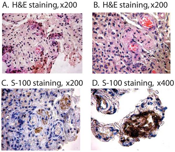
Figure 4.
Teratoma formation from the induced multipotent cells. Skin fibroblasts were exposed to fish occyte extracts and were transplanted subcutaneously into the dorsal flank of nude mice. Two weeks after the injection, tumors were surgically dissected and stained with hematoxylin and eosin (A, B). The presence of neural structures was identified by immunostainign with S-100 antibody (C, D).

