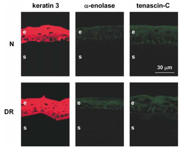
Fig. 3.

Distribution of keratin 3, α-enolase, and tenascin-C in control organ-cultured corneas. In both normal (N) and DR corneas, all markers have patterns very similar to the in vivo central corneas. Keratin 3 is present in all epithelial layers, limbal marker, α-enolase, is scarce to absent, and fibrosis marker, tenascin-C, is absent. These patterns did not change in wounded and healed corneas (not shown here). All corneas were kept in organ culture for 13 days. Central parts of all corneas are shown. E, epithelium; S, stroma. Indirect immunofluorescence.
