
Fig. 4.
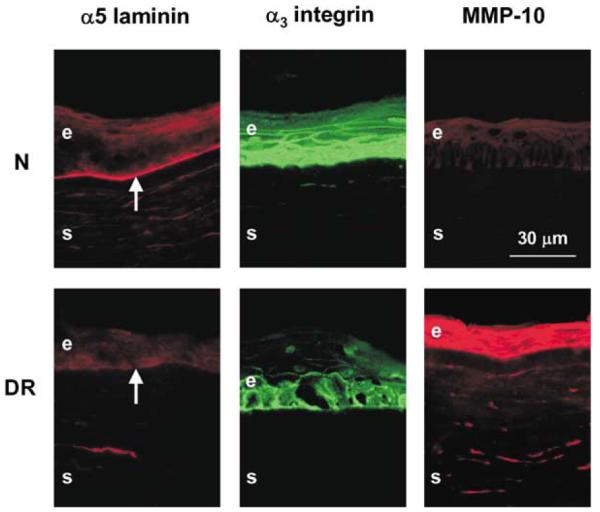
Distribution of DR markers in wounded and healed normal and DR organ-cultured corneas. Note that in both normal (N) and DR corneas, marker distribution is the same as in the respective unwounded corneas (Fig. 2). Normal corneas presented were cultured for 10 days (8 days after healing was complete). DR corneas presented were cultured for 13 days (9–10 days after healing was complete). Central parts of all corneas are shown. E, epithelium; S, stroma; arrows point to the epithelial BM. Indirect immunofluorescence.