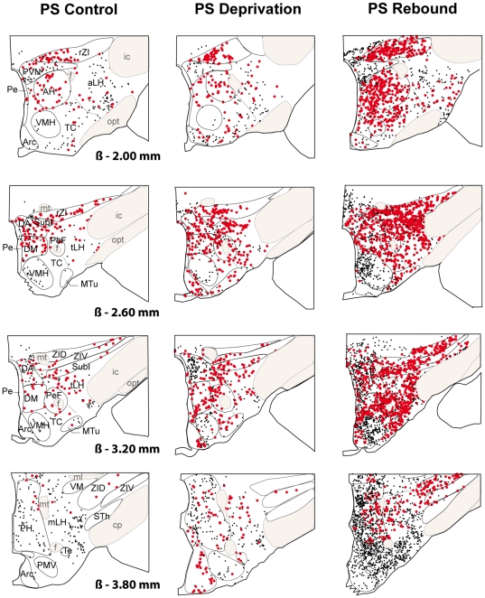
Figure 2. Schematic distribution of Fos-ir (small black dots) and Fos-ir/GAD+ (large red dots) neurons on 4 coronal sections taken at 600µm intervals in a representative animal for PSC (left hand side), PSD (middle) and PSR (right hand side) conditions after Fos immunohistochemistry combined with GAD67 in situ hybridization.
Sections from −2.00 to −3.80 from Bregma (ß). Abbreviations: AH: anterior hypothalamic area; aLH: lateral hypothalamic area, anterior part; Arc: arcuate nucleus; cp: cerebral peduncle; DA: dorsal hypothalamic area; DM: dorsomedial hypothalamic nucleus; f: fornix; ic: internal capsule; ml: medial lemniscus; mLH: lateral hypothalamic area, mammillary part; mt: mammillothalamic tract; opt: optic tract; MTu: medial tuberal nucleus; Pe: periventricular nucleus; PeF: perifornical nucleus; PH: posterior hypothalamic area; PMV: premammillary nucleus, ventral part; PVN: paraventricular hypothalamic nucleus; rZI: zona incerta, rostral part; SubI: subincertal nucleus; STh: subthalamic nucleus; TC: tuber cinereum area; Te: terete hypothalamic nucleus; tLH: lateral hypothalamic area, tuberal part; VM: ventromedial thalamic nucleus; VMH: ventromedial hypothalamic nucleus; ZID: zona incerta, dorsal part; ZIV: zona incerta, ventral part.

