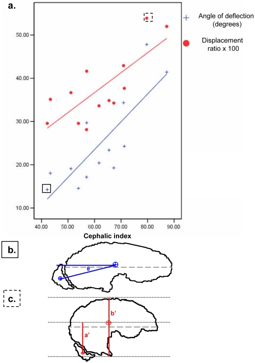
Figure 3. Deviation of the Olfactory Lobe.
a) Cephalic index and deviation of the olfactory lobe using two different methods after normalization of cerebral axis to horizontal. Two exemplar dogs highlighted in boxes are illustrated in parts b) and c) below. b) Angle of deflection method: angle in degrees between centre of mass of brain (CoMbrain) and centre of mass of olfactory lobe (CoMOL). c) Displacement method: Ratio of the ventral-dorsal distance from centre of mass of brain and centre of mass of olfactory lobe (a′) to overall brain height (b′).

