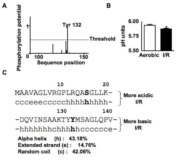
Figure 7.
Identification of putative kinases for the phosphorylation of the βE1 of PDH. (A) Graph showing the phosphorylation potential of amino acids in the more basic form of the βE1 of PDH. (B) Isoelectric point of the more basic form of the βE1 of PDH. (C) Predicted secondary structure surrounding the putative phosphorylation sites.

