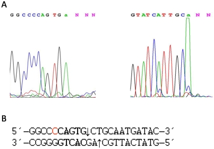
Figure 4. Run-off sequencing to determine the actual cutting site of BtsI-1.
A: pUC19 (0.6 µg) was digested by BtsI-1(∼1.8 pmol) and subjected to run-off sequencing with two flanking primers for both directions. The drop in the peak reflects where the polymerase runs off the template at the nicked sites. The terminal “a” is automatically added to the end by the polymerase during sequencing. B: The sequencing data were aligned with pUC19 sequence. The nicking sites are indicated by arrows on both strands.

