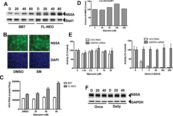
Figure 3. Silymarin does not block HCV replication in HCV replicon cell lines.
Cells were treated once with the indicated doses of silymarin and incubated for 72 hours before replication was assessed. A, protein expression in subgenomic BB7 and genomic FL-NEO HCV-1b replicons following treatment with 20–80 μM of silymarin. D=DMSO. Positions of HCV NS5A and cellular Stat1 proteins are indicated. B, Intracellular NS5A protein expression in DMSO and silymarin treated FL-NEO cells. Cells were treated as described above and NS5A protein detected by immunofluorescence as described in the Materials and Methods. NS5A positive cells are depicted in green while the blue cells represent nuclei counterstained by DAPI. C, HCV RNA levels in FL-NEO and BB7 cells. D, Effect of silymarin against Luc-ubi-neo/ET cells, a Con1-based genotype 1b replicon subgenomic replicon. Values represent percent change in luciferase light units relative to DMSO control. E, Effect on HCV-1a subgenomic replicon. Left panel shows HCV and GAPDH RNA levels following Silymarin treatment, while right panel shows RNA levels following treatment with Intron-A (recombinant IFN-α). F, Effect of silymarin on subgenomic genotype 2a JFH-1-derived Huh7 replicon cell line. Cells were treated with 20 or 40μM of silymarin. Positions of HCV NS5A and cellular GAPDH proteins are indicated.

