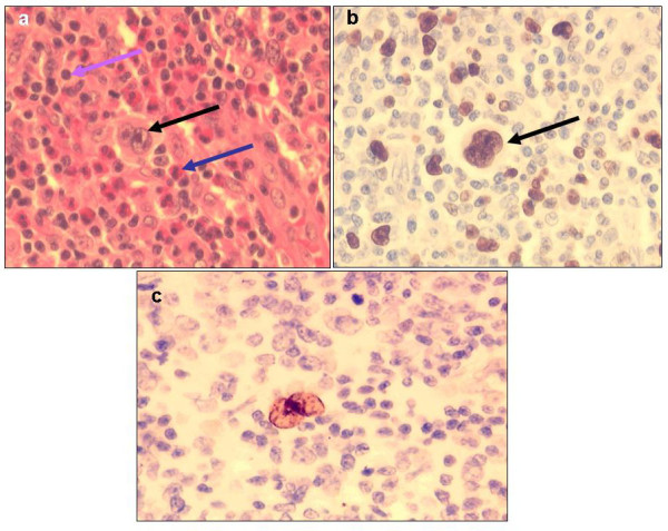
Figure 3.

(a, b, c,)- HL histologic sections: (a) H & E staining of a classical HL mixed cellularity (MC) case showing Reed-Sternberg (RS) cells (black arrow), eosinophils (blue arrow) and lymphocytes (purple arrow), (b) nuclear Ki-67 immunoperoxidase reactivity (brown) in an RS cell, and (c) an EBER positive (brown) RS cell (all ×400).
