
Figure 4.
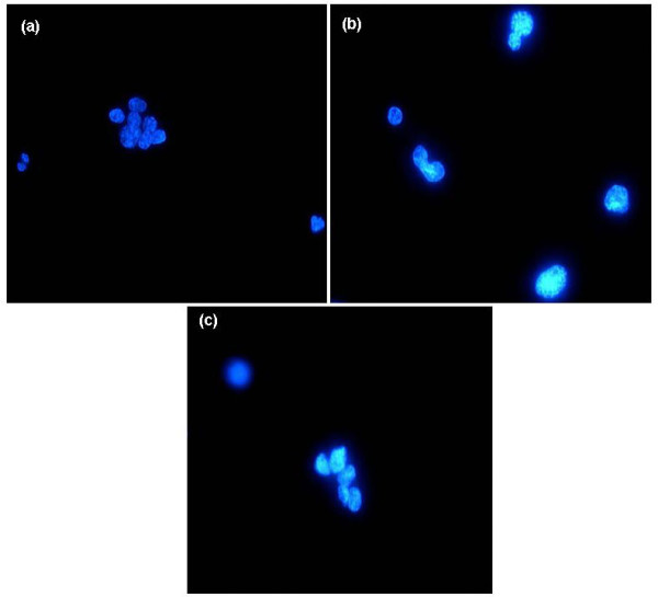
(a, b, c)- DAPI stained nuclei from a (a) normal paraffinized tonsil showing relative monomorphism and from DLBCLs showing pleomorphism (b & c) and mitoses (b).
Official websites use .gov
A
.gov website belongs to an official
government organization in the United States.
Secure .gov websites use HTTPS
A lock (
) or https:// means you've safely
connected to the .gov website. Share sensitive
information only on official, secure websites.
(a, b, c)- DAPI stained nuclei from a (a) normal paraffinized tonsil showing relative monomorphism and from DLBCLs showing pleomorphism (b & c) and mitoses (b).