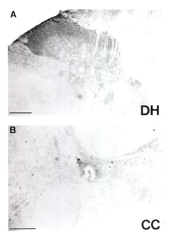
Figure 1.
Light microscopy pictures of the anti-Id mAb immunoreactivity: In (a) at the level of the dorsal horn in cervical segment of the spinal cord, the labelling is particularly intense in laminae I and II as well as at the level of the dorsolateral funiculus. In (b) at the level of the central canal at the same cervical level, the immunoreactivity is mostly localized at the level of the lamina X. Scale bar = 200 μm.

