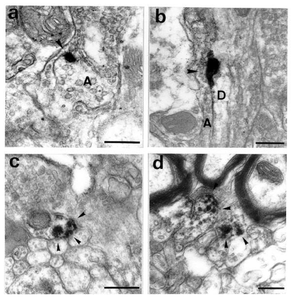
Figure 4.
Immunoreactivity observed in the lamina I of the rat dorsal horn after dermenkephalin stimulation. In (a) and (b) after 15 minutes under dermenkephalin stimulation, labelled axons (A) or dendrites (D) were observed. Dense immunoreactive zones (arrowheads) were associated with invagination of the plasma membrane. In (c) and (d), after 30 minutes stimulation, immunoreactivity was found in the cytoplasm of the neurites. Labelling was associated with large vesicles close to the plasma membrane (c). Labelled microtubules were also found (d). Scale bars = 500 nm.

