Figure 3.
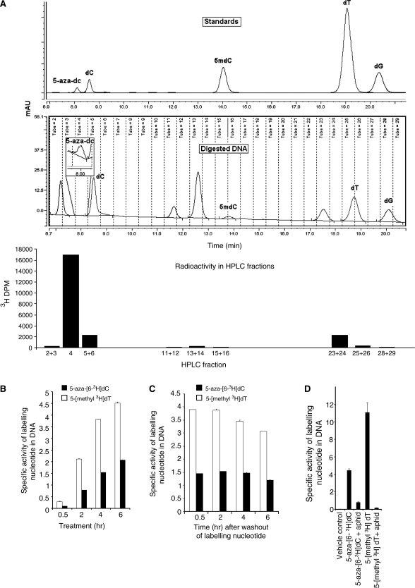
(A) Chromatogram of nucleoside standards (top panel) and nucleosides from digested DNA of cells treated with 5-aza-[63H]-dC for 4-h post-medium replacement (middle panel). The bottom panel shows 3H-DPM of fractions collected in the panel above. Specific activity of DNA from HCT116 cells treated with aphidicolin (20 μg/ml) for 24 h followed by treatment with 5-aza-[63H]-dC and [5-methyl-3H]-thymidine for 12 h. (B) Specific activity of 5-aza-[63H]-dC and [5-Methyl-3H]-thymidine in HCT116 cells treated for 0.5, 2, 4 and 6 h. (C) Specific activities at 0.5, 2, 4 and 6 h after washout of labeling nucleotides. (D) The incorporation of 5-aza-[63H]-dC and [5-methyl-3H]-thymidine into HCT116 cell DNA, with and without prior treatment with the DNA synthesis inhibitor aphidicolin at 20 μg/ml for 24 h.

