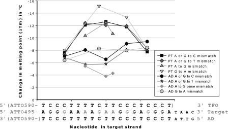
Figure 1.
Introduction of a base mismatch along the target strand and change in melting point (ΔTm) of the AD and the parallel triplex TFO. The ADs consist of the oligonucleotides D-200 to D-222 as target strands with Base mismatches in combination with D-484 as AD strand. The PT consist of the oligonucleotides D-200 to D-222 as target strands with base mismatches in combination with D-256 as AD strand and D-284 as triplex TFO strand. Triplex melting points were determined using 1.0 µM of each strand in sodium acetate buffer at pH 5.0. AD melting points were determined using 0.5 µM of each strand in sodium phosphate buffer at pH 7.0. Base mismatch positions are marked in gray. Fluorophore and TFO strand are placed in brackets as they are not used in all experiments. PT is parallel triplex, AD is antiparallel duplex, A is adenine, G is guanine, C is cytosine and T is thymine.

