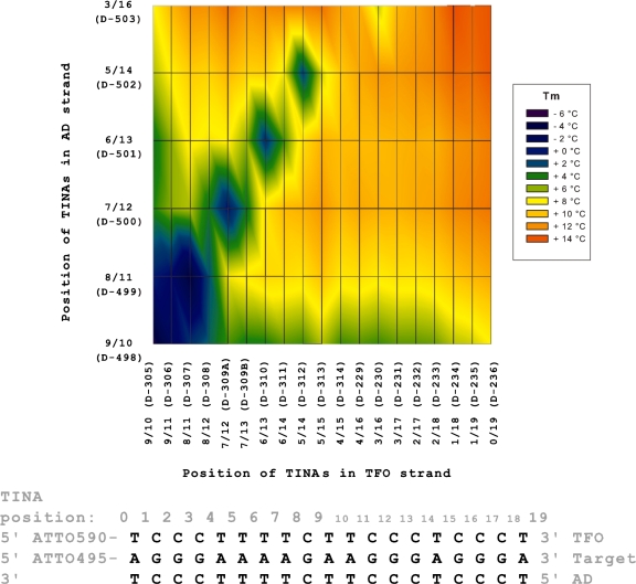
Figure 2.
Position of two TINAs in both the TFO and AD strands of the triplex and change in Tm. Parallel triplex with two TINAs in the AD strand and two TINAs in the TFO strand and change in triplex Tm. On both axes TINAs are moved from the centre to the ends of the oligonucleotide. Triplex Tm was determined using 1.0 µM of each strand in NaOAc-buffer at pH 5.0. TINA positions are indicated above the triplex sequence counted from the 5′ to the 3′ position in the target strand, and the numbers reflect the position of the base 5-prime to the TINA bulge insertion. Colour indicates the change in Tm compared with a pure DNA triplex.

