Figure 2.
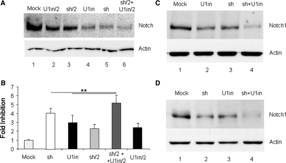
Inhibition of endogenous Notch1 expression and function with RNAi and U1i. (A). Inhibition of endogenous Notch1 expression. HeLa cells were transfected with a control plasmid (mock), with plasmids expressing U1inαNotch1 (U1in) or shαNotch1 (sh) at the highest dose, or half of the highest dose (X/2) or with the combination of plasmids expressing U1inαNotch1 and siαNotch1 (sh/2 + U1in/2). HeLa cell lysates were collected 3 days after transfection and Notch1 (upper box) and actin (lower box) expression was evaluated in two independent western blots. (B). Functional assay for Notch1 gene silencing. 293 cells were co-transfected with pNFκβ-Luc, Renilla luciferase control and the plasmids described in A. Luciferase activity was evaluated 3 days after transfection and the data obtained were corrected for equal transfection efficiency. Results were plotted to indicate the fold inhibition obtained in each case. Fold inhibition was calculated as the ratio of the luciferase activity obtained in mock transfected cells versus the luciferase activity obtained in cells expressing the indicated inhibitor. Error bars denote standard deviations of three independent experiments. (C and D) Analysis of the silencing of endogenous Notch1 over time. (C) Pools of clones of HeLa cells stably transfected with a control plasmid (mock), or plasmids expressing U1inαNotch1(U1in), shαNotch1 (sh) or both (sh + U1in) were used to evaluate Notch1 (upper box) and actin (lower box) expression by western blot. (D) Representative clones of HeLa cells expressing U1WT or U1inαNotch1 were transfected with a control plasmid (mock and U1in, respectively) or with a plasmid expressing shαNotch1 (sh and sh + U1in, respectively). Stably transfected pools of clones were used to determine Notch1 (upper box) or actin (lower box) expression by western blot.

