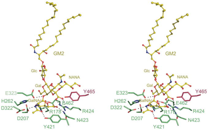
Figure 4.
Model of GM2 docked onto the α-subunit active site of Hex A. A model of the GM2 ganglioside (yellow) was docked into the active site of the α-subunit of Hex A based on the model of GM2 bound to the α-subunit active site of Hex B.18 For clarity, only residues interacting with the sugar residues of GM2 are shown. GM2AP, which interacts with the acyl chains of the GM2 ganglioside, has also been removed. αArg424, a positively charged residue unique to the α-subunit of Hex A, is found within hydrogen bonding distance from the negatively charged carboxylate of the NANA group of GM2.

