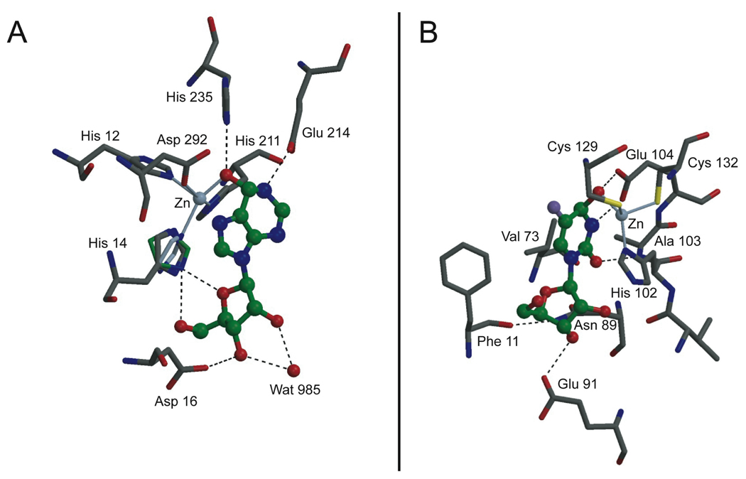
Figure 2.
(A) Bovine ADA in complex with hydrated nebularine.[11] Ligand is shown in ball-and-stick representation, with green carbons. The crystal coordinates of residue His 14 are shown in dark gray. This conformation is suggested to be an average of two rotamers: one (in green) where the Nδ atom makes a bifurcated hydrogen bond to the sugar moiety in the ligand, and one (in light blue-gray) where the zinc atom lies in the plane of the imidazole ring. (B) E. coli CDA in complex with hydrated 5F-zebularine.[13]

