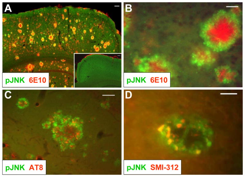
Figure 1. JNK is activated in dystrophic neurons in the peri-plaque region in Tg2576/PS1M146L mice.

(A) 12-month old Tg2576/PS1M146L transgenic mice show strong 6E10 immunoreactivity indicative of Aβ deposition (red) and increased JNK phosphorylation (green) in the cortex compared to wild-type mice (inset). Scale bar, 100 μm. (B) At higher magnification, phosphorylated JNK (green) is seen to surround amyloid plaques (red) in cortex. Scale bar, 20 μm. (C) JNK phosphorylation (green) partially overlaps with immunoreactivity with the AT8 antibody, indicative of hyperphosphorylated tau. Scale bar, 50 μm. (D) JNK phosphorylation (green) also coincides with SMI-312 immunoreactivity, indicative of dystrophic neurons. Scale bar, 20 μm. All images are representative of observations from at least 3 animals.
