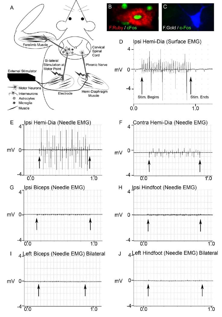
Figure 1. Phrenic nerve hyperstimulation: technique development in SOD1G93A rats.

The diagram illustrates the phrenic nerve motor endpoint stimulation technique (A). Stimulating electrodes were bilaterally implanted into the center of both hemi-diaphragm muscles to achieve phrenic nerve activation upon external stimulation. Even though placed directly into muscle, the electrode ends were positioned near the phrenic nerve motor endpoints to allow for stimulation of the phrenic nerve (stimulation/contraction is unsuccessful if there is not phrenic nerve innervation of the diaphragm). The stimulator is found externally, and the ends of the 2 electrodes are connected to the stimulator each day during stimulation, while the animal is awake and freely-behaving. Motor neurons innervating the diaphragm originate in the cervical spinal cord, in regions where forelimb motor neurons also reside. This paradigm allows for the study of focal effects on the peripheral nervous system (muscle and nerve), as well as central nervous system effects on motor neurons, astrocytes, and microglia - all key cell types in mutant SOD1-mediated disease (A).
Following bilateral phrenic nerve stimulation, c-Fos expression was detected in fluororuby retrogradely-labeled phrenic motor neurons (Fig 1B), but not in fluorogold-labeled forelimb motor neurons (Fig 1C), suggesting that phrenic nerve stimulation activated phrenic motor neurons, but not forelimb motor neurons. To also examine whether external diaphragm stimulation resulted in ectopic stimulation of other motor neuron / muscle groups (besides phrenic nerve / diaphragm), a comprehensive series of intra-muscular needle EMG and muscle surface EMG recordings were obtained during unilateral phrenic nerve stimulation of SOD1G93A rats. Stimulation resulted in consistent surface electrical responses in the ipsilateral diaphragm lasting the duration of the programmed stimulus duration (D). Robust intramuscular electrical activity (∼3.5mV) was obtained in the ipsilateral hemi-diaphragm (E) with needle EMG recordings, while a smaller response (∼1.0mV) was also noted in the hemi-diaphragm contralateral to stimulation (F). On the contrary, significant electrical responses during phrenic nerve stimulation were not found with intra-muscular needle EMG recordings in the ipsilateral biceps (G) or ipsilateral hindfoot (H). Needle EMG recordings were also conducted in the leftt biceps (Fig 1I) and left hindfoot (Fig 1J) during bilateral phrenic nerve stimulation, and significant electrical activity was not observed. Arrows denote beginning and end of stimulation.
