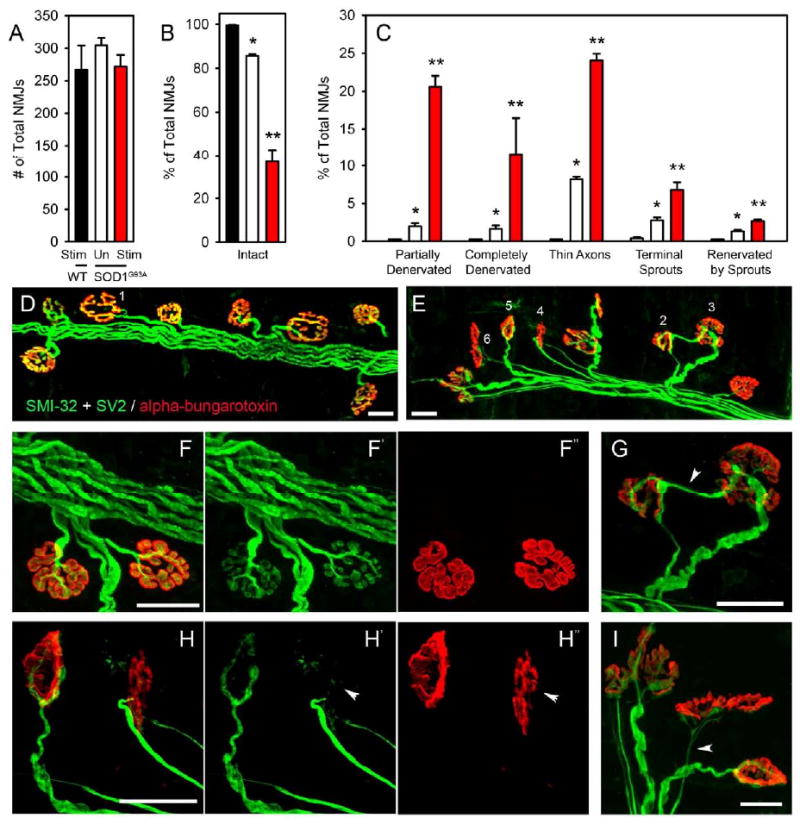
Figure 5. Phrenic nerve hyperstimulation stimulation accelerated pathological changes at diaphragm neuromuscular junctions in SOD1G93A rats.

There were no differences in total numbers of NMJs among any groups, as assessed by total numbers of alpha-bungarotoxin+ junctions (A). Nearly all wild-type NMJs were completely intact, characterized by: complete overlap of the pre-synaptic axon and pre-synaptic vesicles with post-synaptic acetylcholine receptors, no signs of multiple innervation, absence of pre-synaptic axon thinning (D: NMJ #1, F). While both groups of SOD1G93A rats showed signs of synaptic disruption, stimulated SOD1G93A rats had significantly fewer intact junctions, with greater than 60% of junctions being affected (B, E). To specifically examine the types of changes occurring in stimulated diaphragm muscle, NMJ changes were broken down into a number of phenotypic categories. Compared to unstimulated SOD1G93A rats, a significantly greater percentage of junctions from stimulated SOD1G93A rats showed signs of partial denervation (C, E: NMJ #4 and #5, H - arrowhead), complete denervation (C), thinning of the pre-synaptic axon (C, E: NMJ #6, I -arrowhead), terminal sprouting (C, E: NMJ #3, G - arrowhead) and reinnervation of denervated junctions by terminal sprouts from adjacent junctions (C, E: NMJ #2, G). In many instances, individual junctions showed signs of more than 1 change. Only rare instances of any of these junctional pathologies were found in wild-type stimulated muscles. Scale bars: 25μm.
