
Figure 6.
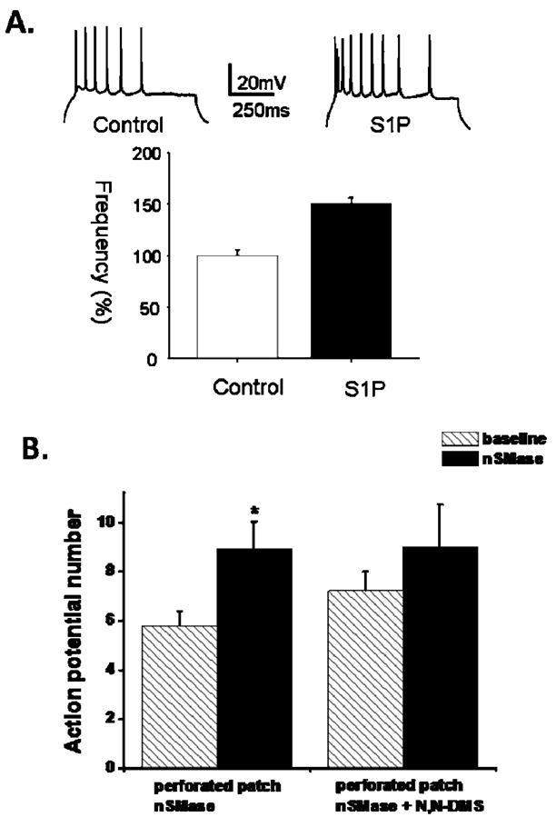
Evidence that S1P generation is sufficient and necessary for the increased neuronal excitability resulting from nSMase activation. A. S1P increases action potential firing frequency in CA1 pyramidal neurons. Upper panel): Representative traces generated from a single 50 pA depolarizing step measured 15 minutes after establishing whole cell condition in the absence (control) and presence (S1P) of S1P in the intracellular pipette. Lower panel): Cumulative data (bottom panel) showed a significant difference in the firing frequency between control (n = 8) and S1P-treated (n = 8) neurons. B. nSMase-mediated increase in action potential firing is attenuated by sphingosine-kinase inhibition. Action potential firing under perforated patch condition was increased following nSMase exposure (15 min; 0.4 U/ml) compared to baseline (n = 10). Incubation of slices with the sphingosine kinase inhibitor N,N-DMS (10 uM) for 30 – 90 min prior to nSMase application (15 min; 0.4 U/ml, n = 5) blocked the increase in action potential firing. Data are presented as the mean ± SEM. Student’s t-test was used for statistical comparisons. *p < 0.05.