
Figure 1. Expression of gp120 in 293T cells results in a mixture of monomeric and dimeric gp120.
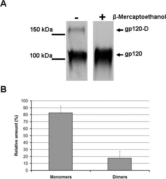
(A) A transfected 293T cell supernatant containing radiolabeled wild-type gp120 was incubated with a serum mixture from HIV-1-infected individuals for two hours at 37°C. Precipitates were analyzed by SDS-PAGE in the presence or absence of 5 mM β-mercaptoethanol followed by autoradiography/ densitometry. The result shown is representative of those obtained in at least three independent experiments. The gp120 dimer is designated gp120-D. (B) The bands presented in (A) were quantified by densitometry. Results are expressed as the percentage of each different form (monomers or dimers) relative to the total amount of gp120 (monomers + dimers). Data shown represent the means +/- SEM of two independent experiments.