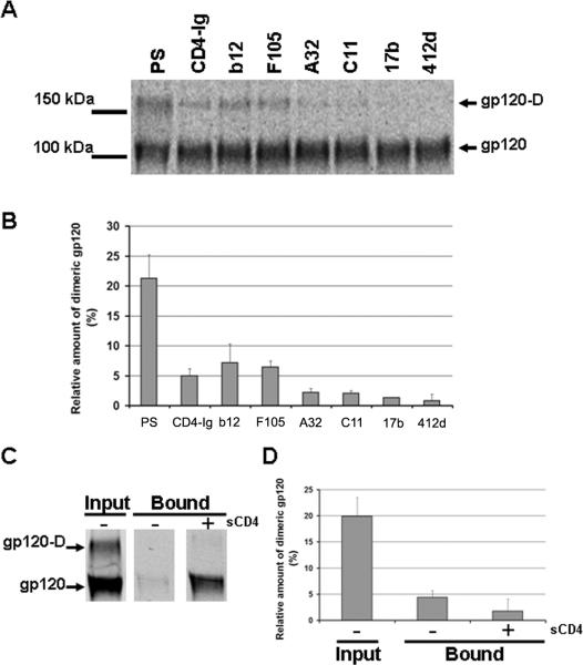
Figure 2. Dimeric gp120 is poorly recognized by CD4-induced antibodies and CCR5.
(A) A 293T cell supernatant containing radiolabeled wild-type gp120 was incubated with a polyclonal mixture of sera from HIV-1-infected individuals (PS) or 13 nM of monoclonal antibodies or CD4-Ig for two hours at 37°C. Precipitates were analyzed by SDS-PAGE without β-mercaptoethanol followed by autoradiography/densitometry. The result shown is representative of those obtained in two independent experiments. The gp120 dimer is designated gp120-D. (B) The gp120 bands detected in (A) were quantified by densitometry. Results are expressed as the percentage of dimeric gp120 relative to the total amount of gp120 (monomers + dimers). Data shown represent the means +/-SEM of two independent experiments. (C) Radiolabeled HIV-1YU2 gp120 glycoproteins in 293T cell supernatants were incubated in the absence or presence of 200 nM sCD4 prior to addition to Cf2Th cells expressing CCR5. After two hours at 37°C, the amount of input and bound gp120 was determined by immunoprecipitation with PS. Samples were analyzed by SDS-PAGE under non-reducing conditions. The gp120 dimer is designated gp120-D. (D) The gp120 bands detected in the experiment shown in (C) were quantified by densitometry. The percentage of dimeric gp120 in each sample relative to the total gp120 is shown. The data represent the means +/- SEM of two independent experiments.

