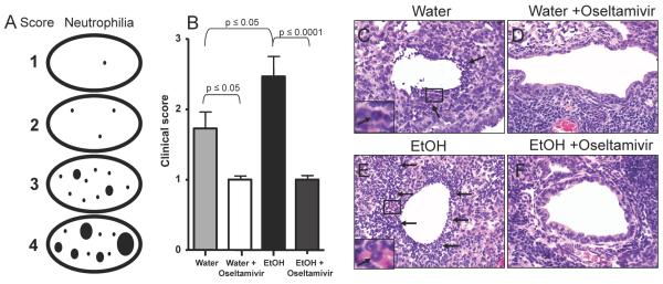
Fig. 4.
IAV induced neutrophilia is decreased in oseltamivir treated chronic EtOH exposed mice. A.) Schematic of scoring for neutrophilia in low magnification lung sections (ovals). Black dots represent lesion distribution with increased dot size representing increased severity (see Materials and Methods for further description of the scores). B.) Mice were infected and treated with oseltamivir as described in Fig. 1A. On day 14 p.i. pulmonary neutrophilia was measured. Values represent the clinical score assigned by a blinded Veterinary Pathologist. Data are pooled from 2 independent experiments with 5-9 mice per group. C.) Representative sample of a control water mouse receiving no treatment showing minimal levels of neutrophilic infiltration. D.) Representative sample of an untreated EtOH mouse with enhanced neutrophilic foci in the airway. E.) Representative sample of a water mouse receiving oseltamivir treatment showing reduced levels of neutrophilic infiltration. F.) Representative sample of an EtOH mouse receiving oseltamivir treatment showing a significant reduction in airway neutrophilia compared to untreated EtOH mice. Arrows indicate areas of neutrophilic infiltration. All images shown are at 40x magnification. Data are representative of 2 independent experiments with 5-9 mice per group.

