
Figure 5.
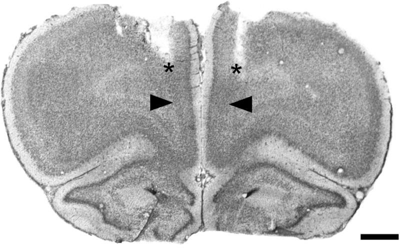
Representative photomicrograph of a Cresyl Violet-stained coronal section through mPFC, corresponding to plate 9 in the atlas of Paxinos and Watson (1998), showing the tracks of implanted guide cannulae (asterisks), and the microinjection sites, located 1 mm below the guide cannulae (arrowheads). See fig. 2 for region labels. Scale bar = 1 mm.