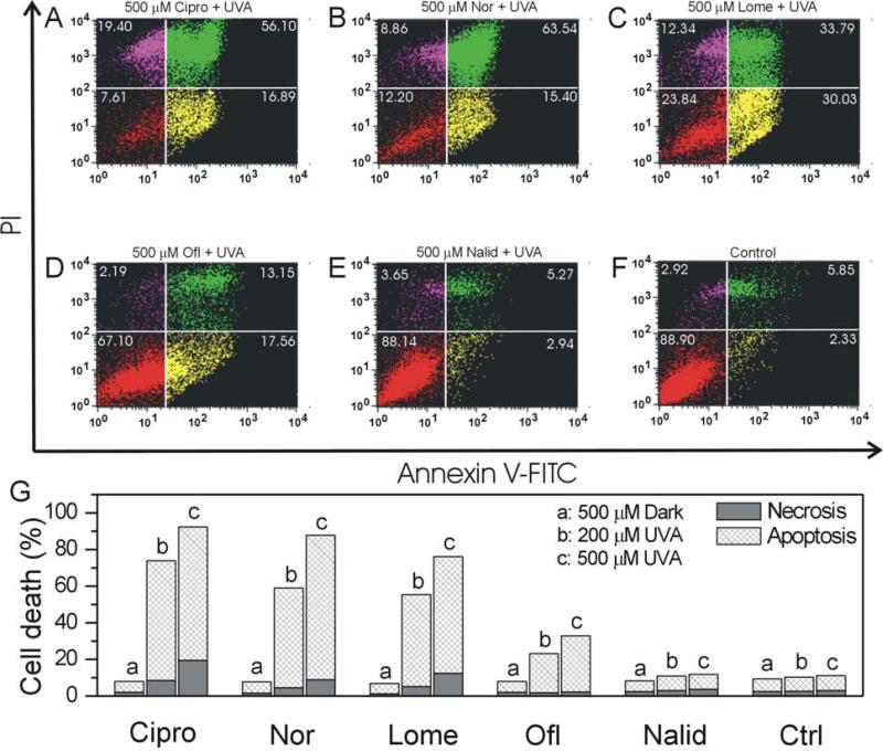
Figure 7.
FLQ/quinolone-induced apoptotic and necrotic death in HLE B-3 cells with UVA radiation (A: Cipro, B: Nor, C: Lome, D: Ofl, E: Nalid, and F: Control). Cells were seeded in plastic petri dishes (60 cm2) and pretreated with different FLQs at 500 μM concentration in HBSS for 1 hour and then exposed to UVA (10 min). After radiation, the cells were incubated overnight in cell culture medium and then stained with annexin V-FITC and propidium iodide. Apoptotic and necrotic cell death were determined with flow cytometry. The lower left quadrant shows normal viable cells, the lower right and upper right quadrants show apoptotic cells, while the upper left quadrant shows necrotic cells. (G) Graphs illustrating apoptosis and necrosis induced by UVA with different FLQs/quinolone.

