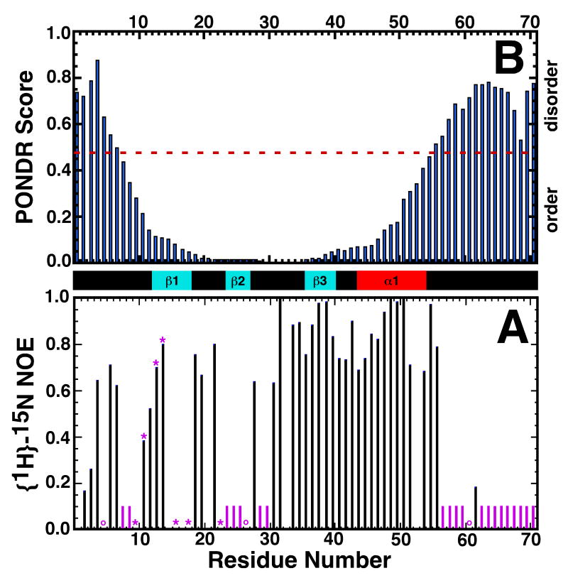
Figure 4.
A) Backbone {1H}-15N heteronuclear NOE values for Rv2377c. The position of proline residues in the sequence are indicated by purple circles. Amide cross peaks that were absent or very weak in the original 1H-15N HSQC spectrum are indicated by purple bars and asterisks, respectively. B) Graphical output of a PONDR prediction on Rv2377c using the VL-XT algorithm. Consecutive values above and below 0.5 predict disordered and ordered regions, respectively, within the protein. The experimentally observed elements of secondary structure are shown between the two plots: cyan = β-strand, red = α-helix.

