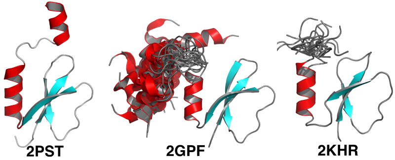
Figure 6.
Comparison of the structure of P. aeruginosa PA2412 determined by XRD (2PST) and NMR (2GPF) methods with the NMR determined structure of Rv2377c (2KHR). The structure in the NMR ensemble closest to the average structure is shown. The position of the second α-helix in 2GPF, relative to the structured core, is shown for each of the 20 structures in the PDB deposited ensemble. Similarly, a short part of the C-terminal region for the PDB deposited ensemble for 2KHR is also shown. In both cases the extreme N- and C-terminal regions for 2GPF and 2KHR have been removed for clarity, but, do include the highly conserved region, WXDXR. The β-strands are colored blue and the α-helices are colored red.

