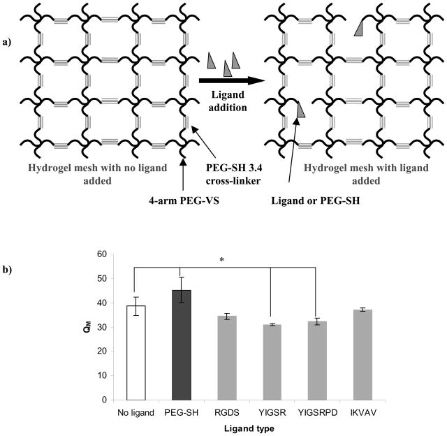
Figure 3.
a) Schematic representation of hydrogel mesh disruption upon covalent binding of ligand or PEG-SH to the 4-arm PEG-VS; b) Influence of ligand type on PEG hydrogel swelling ratio (QM). All hydrogels were prepared as 10% w/v polymer with 100 μM ligand. Asterisks designate significant differences.

