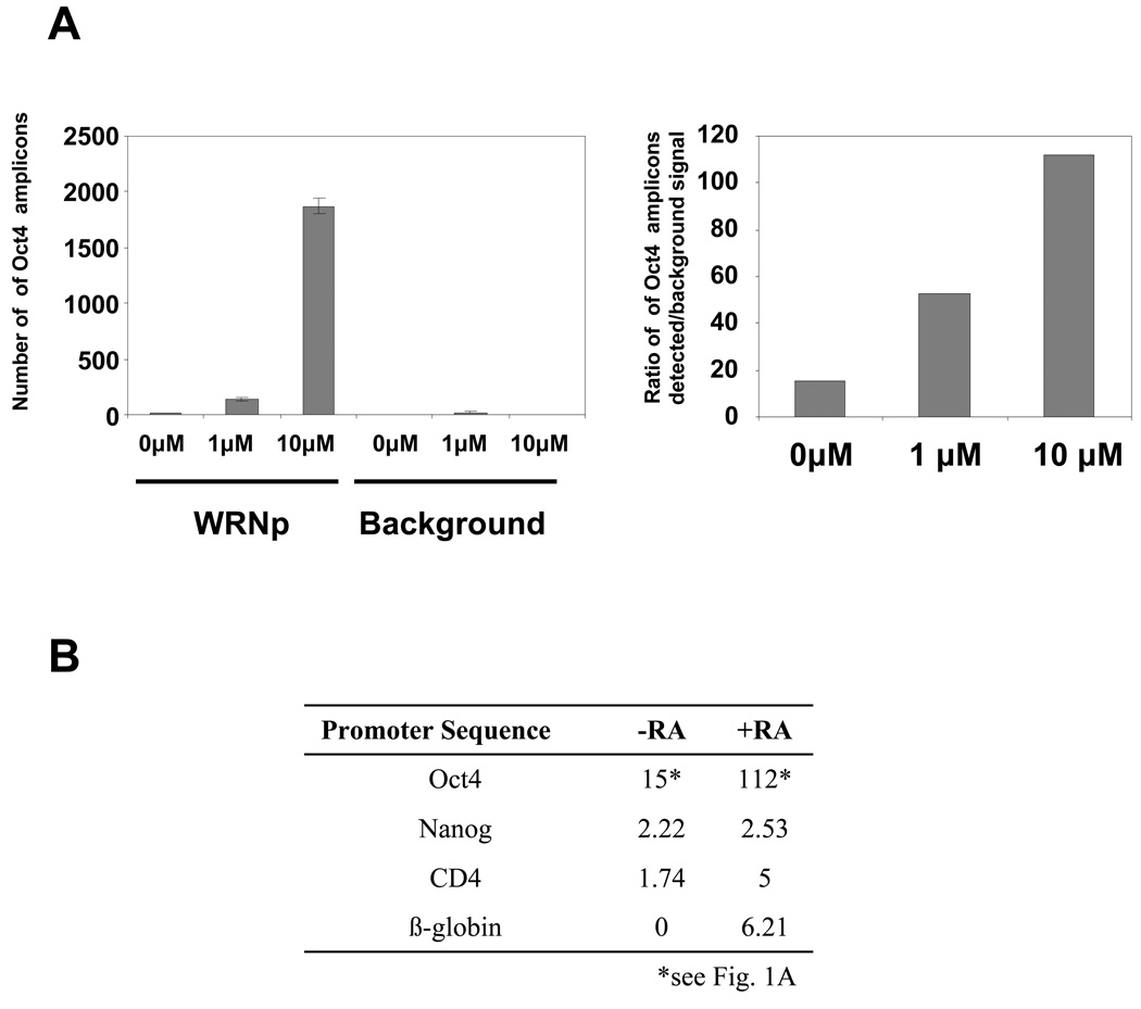
Fig. 1. Enrichment of WRNp at the Oct4 promoter.
(A) Q-ChIP analysis of WRNp levels at the Oct4 promoter in undifferentiated pluripotent cells and cells stimulated to differentiate with either 1 or 10 µM RA for three days. WRNp was immunoprecipitated from NCCIT lysates and the amount of associated Oct4 promoter DNA was measured by real-time PCR using the Oct4 B probe and primers (See Supplementary Table 2). Left – raw data, Oct4 promoter amplicons detected in samples with 0, 1, or 10 µM RA, immunoprecipitated with a WRN antibody (WRNp), or without antibody to determine extent of background signal (Background). Error bars indicate standard deviation. Right – ratio of Oct4 promoter amplicons detected in lysates immunoprecipitated with the antibody over the signal detected in no antibody control samples. (B) WRNp enrichment at the Nanog, CD4 and β-globin promoters in the presence and absence of RA (fold enrichment over background). Cells were treated as in A.

