
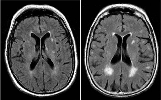
Fig 1 White matter hyperintensities on magnetic resonance imaging (axial fluid attenuated inversion recovery sequence) in two 80 year old patients: (left) minor white matter hyperintensities; (right) extensive white matter hyperintensities predominating in periventricular region. White matter lesions are considered present if hyperintense on T2 weighted, fluid attenuated inversion recovery, and proton density images, without prominent hypointensity on T1 weighted images