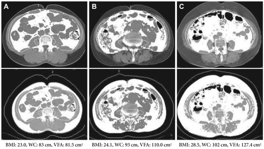
Fig. 3.
Images demonstrate our method for determining abdominal fat distribution on a CT scan obtained at the umbilicus. The white areas demonstrated on upper images were regarded as visceral fat tissue, and the white areas on lower images were regarded as total fat tissue (visceral fat and subcutaneous fat). (A) is the CT scan in patient with normal BMI and normal VFA, (B) in patient with normal BMI and central obesity, and (C) in patient with increased BMI and central obesity. BMI: body mass index, WC: waist circumference, VFA: visceral fat area.

