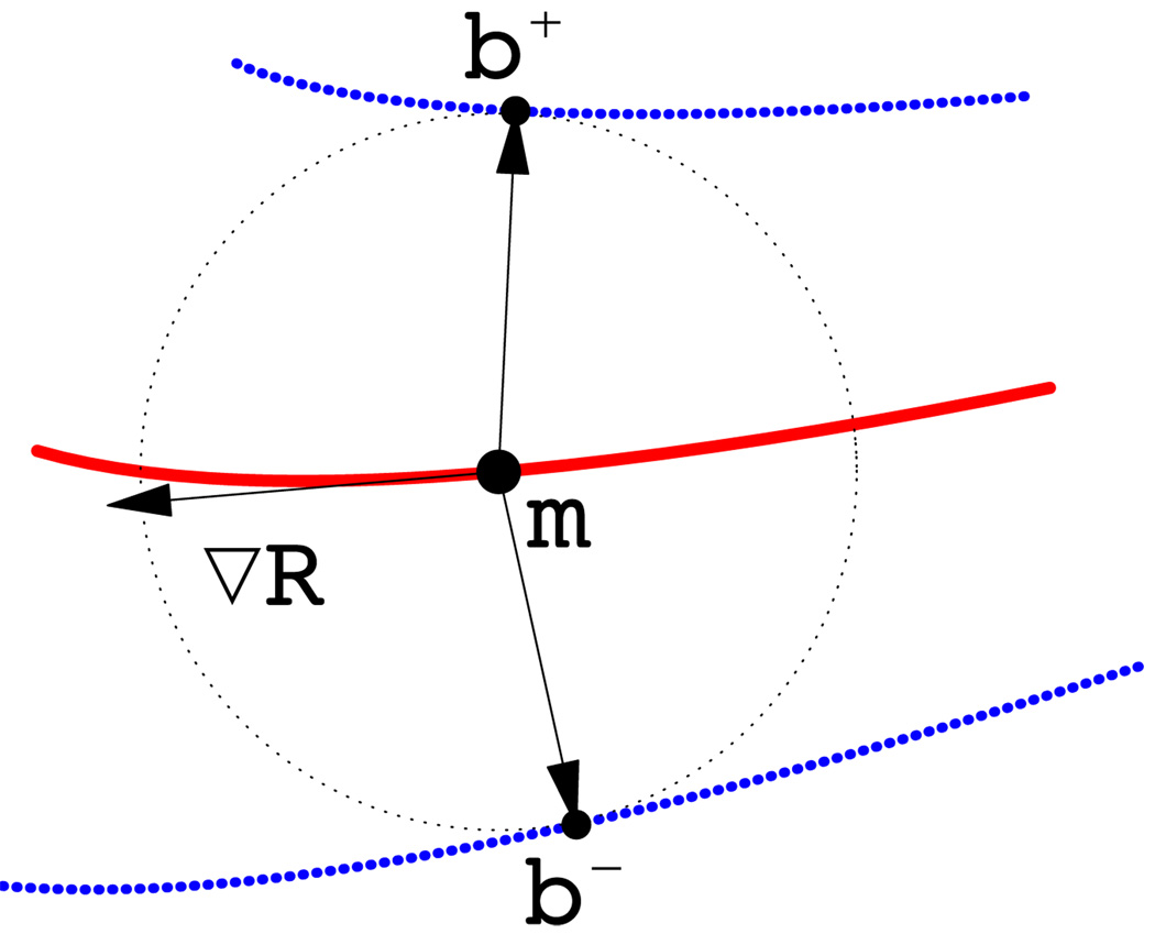
Figure 1.
Two-dimensional diagram of medial geometry. The red curve represents the medial surface (skeleton) m. The circle has radius R, given by the radial field on m. The boundary, shown in blue, consists of two parts, b+ and b−, derived from the skeleton and radial field by inverse skeletonization (Yushkevich et al., 2006). The vector ∇mR lies in the tangent plane of m and points in the direction of greatest change in R. The arrows pointing from m to b+ and b− are called spokes.

