
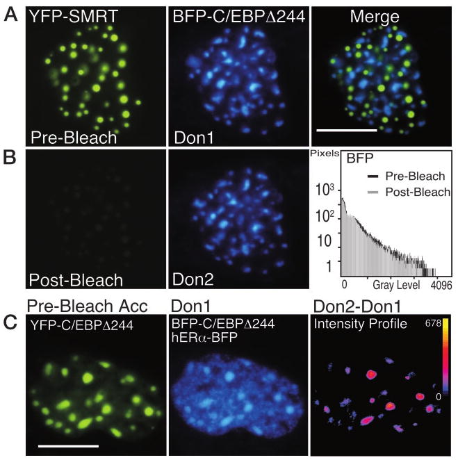
Fig. 4. The Photobleaching of YFP Is Selective.
A, Pituitary GHFT1–5 cells were cotransfected with plasmids encoding the corepressor protein SMRT, tagged with YFP, and the C/EBPΔ244 deletion mutant fused to BFP. Sequential images of the nucleus of a cell coexpressing the proteins were acquired at the same focal plane using the filters described in Materials and Methods. The calibration bar indicates 10 μm. The images were merged to show that the proteins were each localized to discrete subnuclear foci. B, The YFP-SMRT was photobleached by 5 min of exposure to 500-nm light. Postbleach images were acquired at the same focal plane and under identical conditions to document the bleaching of YFP and to show that this had no effect on the BFP signal (histogram). C, Acceptor photobleaching FRET measurements detect only specific protein associations. Pituitary GHFT1–5 cells were cotransfected with plasmids encoding both YFP- and BFP-C/EBPΔ244, and hERα-BFP, and cells were identified that expressed all three labeled proteins. The prebleach images of the acceptor, YFP-C/EBPΔ244 (left panel), and the combined fluorescence from the donor, BFP-C/EBP Δ244 (foci) and the nucleoplasmic-localized hERα-BFP (Don1) are shown. The YFP fluorophore was bleached by greater than 90%, and a second donor image (Don2) was acquired at the same focal plane and under identical conditions as the first. The pixel-by-pixel change in gray-level intensity of the donor signal was obtained by digital subtraction of the Don1 image from the Don2 image. The intensity profile (right panel) represents the change in the gray-level intensities in the dequenched donor image (Don2 − Don1). The calibration bar shows the range of gray-level intensities in the dequenched image with black indicating 0 and yellow indicating the maximum gray-level value (shown next to the calibration bar). Note that the increase in donor intensity was limited to the foci.