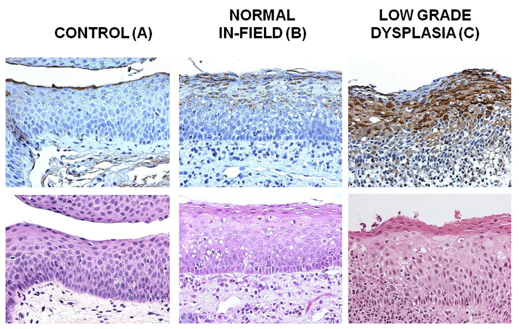
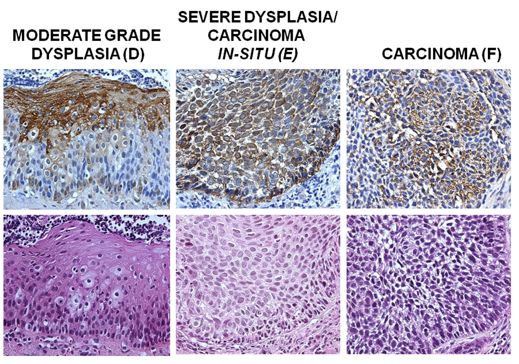
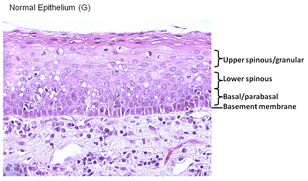
Figure 1. COX-2 staining in premalignant and HNSCC samples.


Tissue blocks were stained with anti-COX-2 specific antibody at a dilution of 1/100 following a standard IHC procedure. Antibody staining was visualized using DAB peroxidase substrate solution and cell nuclei were counterstained using hematoxylin QS. Mouse IgG without the primary antibody was used as a negative control. Representative samples of COX-2 staining in Control tissue (A), Normal in field (B), Low Grade Dysplasia (C), Moderate Grade Dysplasia (D), Carcinoma in Situ (E) is shown. A Representative image is showing the epithelial layers (basement membrane, basal/parabasal layer, lower spinous layer, and upper spinous layer) in a normal epithelial tissue sample, stained with hematoxylin (F).
