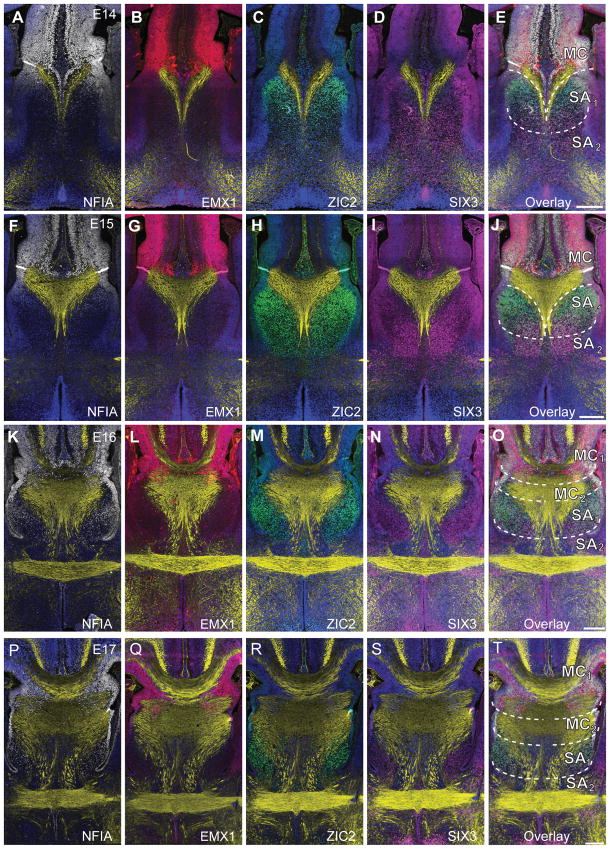
Figure 4.
Molecular patterning and morphology of the developing mouse commissural plate demonstrates the emergence of the four subdomains. Fluorescence immunohistochemistry for the markers NFIA (white), EMX1 (red), SIX3 (magenta) and ZIC2 (green) throughout the developmental oblique coronal plane of the commissural plate in mouse embryos at E14 (A-E), E15 (F-J), E16 (K-O) and E17 (P-T). GAP43 staining is shown in yellow. DAPI counterstain is shown in blue. An overlay of this molecular patterning is shown with boundaries indicated by dashed lines, defining dorsal domains (MC) and ventral domains (SA), characterized as follows: MC or MC1, NFIA+ and EMX1+; MC2 NFIA+ and ZIC2+; SA1, NFIA+ and ZIC2+ and SIX3+; SA2, SIX3+. Scale bar = 200 μm.

