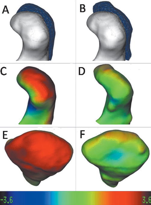
Fig 4.

Left condyle of a patient that was displaced with surgery and showed a stable position with bone remodeling at the 1-year follow-up. A and B, Lateral view of mesh-transparency visualizations of condylar position and morphology: A, superimposed models presurgery (white) and at splint removal (semitransparent mesh; B, superimposed models presurgery (white) and at 1 year postsurgery (semitransparent mesh). Note the stability of the condylar position at 1 year postsurgery in B compared with splint removal in A, but the postero-superior surface of the condyle was flattened in B. C–F, Surface distance color maps visualization: C, lateral view and E, posterior view of condylar models at splint removal displaying the surface distances between presurgery to splint removal; D, lateral view and F, posterior view of condylar models at the 1-year follow-up displaying the surface distances between splint removal and the 1-year follow-up. Note, in more detail in these views, how the postero-superior surface of the condyle was flattened when we compare the models in C and E with the D and F views. Color map ranges between −3.6 mm (dark blue) and +3.6 mm (dark red).
