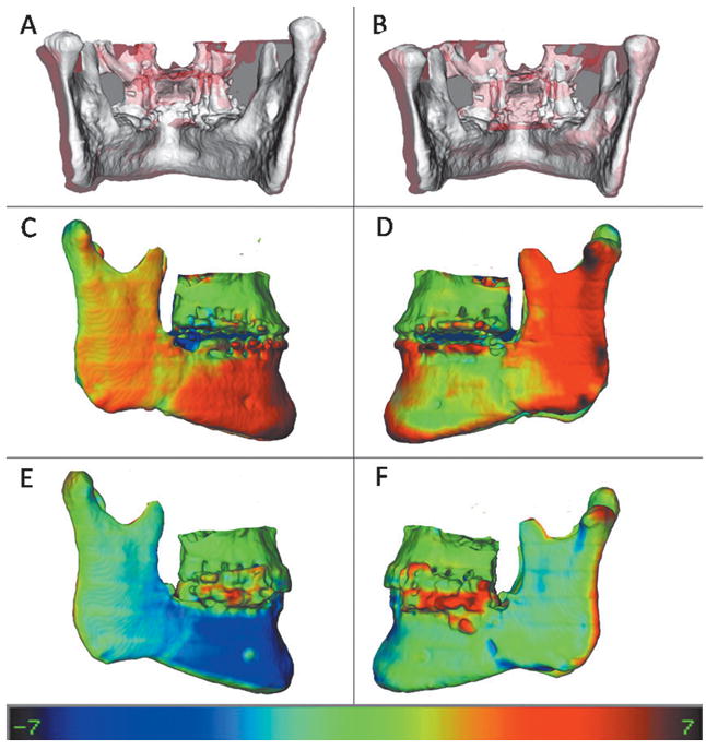
Fig 7.

Example of a patient who had lateral displacement of the rami with surgery on both sides: A, Presurgery is shown in white, and splint removal is shown in semitransparent red; B, presurgery shown in white and 1-year follow-up model is shown in semitransparent red. C and D, right and left views, respectively, of the splint-removal model, displaying the color maps of the surface distances between presurgery and splint removal. E and F, right and left views, respectively, of the 1-year follow-up model, with color maps of the surface distances between splint removal and the 1-year follow-up. The rami movement was maintained at the 1-year follow-up on the left side (F) and relapsed medially on the right (E). Also, note some relapse of chin advancement (blue). Color map ranges between −7.0 mm (dark blue) and +7.0 mm (dark red).
